As the Covid-19 continues to rage the world, the spotlight is back on Dr. June Almeida, a Scottish virologist who pioneered new methods for viral imaging and diagnosis and discovered the first Human Coronavirus.
Born as June Dalziel Hart, in 1930, daughter-of a Scottish bus driver in Glasgow, she was exceptionally bright and ambitious, but had to drop at at the age of 17 for the lack of fundings for university education. But that didn’t deter her from her commitment towards her goal. On leaving school, June became a laboratory technician in hiatoplasty at Glasgow Royal Infirmary and later got a job at St Bartholomew’s Hospital, London in the same discipline. She worked there till 1954.
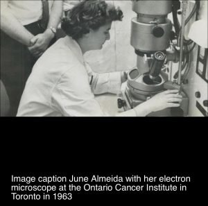
By the time she got married to a Venezuelan artist Enrique Almeida and they moved to Canada. She was hired for the newly opened position of as the electron microscopy technician at the Ontario cancer Institute, where she worked for the next 10 years. With her interest, knowledge and her skills she soon pioneered in her field despite having no formal qualifications. She co-authored numerous scientific publications mostly relating to the structure of virus which until now couldn’t be visualised.
 The microscopic technique Dr. Almeida developed was simple yet revolutionary. She pioneered the Immune Electron Microscopy (IEM) to better image Virus. It included negative staining which involved introducing viruses with specific antibodies raised in animals or human sources, to which the virus then clumped around, which she was able to visualise and ascertain.
It was her IEM technique which led her to be the first person to visualise Rubella even though it was determined to be the cause of congenital malformations if acquired in pregnancy over 25 years previously. Though simple yet revolutionary technique of IEM involving negative staining led to many important discoveries in virology. One of the most notable ones being the presence of two distinct components to the Hepatitis B virus, one in the surface of the particle and another internally. June technique also enabled workers at NIH to visualise the hepatitis A virus for the first time.
In 1964, Professor Tony Waterson who had just been appointed as chair of Microbiology at St. Thomas met Dr. June Almeida while visiting Toronto and offered her to join her research team. She accepted and moved to London. Three years later she joined Royal Post Graduate Medical School with Professor Waterson. By this time Her Publications in her field earned her Doctor of science (D.sc). Her electron micrograph continues to be the basis of most virology review articles and textbooks.
In 1964, Prof. Waterson & Dr Almeida collaborated with Dr. David Tyrrell who led research at the Common Cold Unit (CCU), Salisbury and was deputy director of the Clinical research Centre (CRC), Harrow, Middlesex.
Tyrrell in his book ‘Cold Wars’: The Fight Against The Common Cold (2002) wrote, in 1961 his research team infected Human Volunteers with nasal washing la from pupils at a boy’s boarding school in Surrey, all of whom had common colds. One agent B814, didn’t grow in cells culture but volunteer studies demonstrated its growth in trachea-derived organ cultures and Dr Tyrrell wondered if it could be seen by and electron microscope.
The samples were sent to Dr. June Almeida who “duly dabbed the surface of the tissue on to her microscope grids and examined each one and saw Virus Particles in the B814 specimen which she describes as being rather like influenza virus but not exactly the same.”
In fact, she had seen particles like this earlier while investigating mouse hepatitis and infectious bronchitis of chickens but her paper to a peer – reviews journal was rejected “because the referees said the images she produced were just bad pictures of influenza virus particles.
The specimen would become to be known as the first Human Coronavirus. The discovery was published in the British Medical Journal in 1965. The first photos of Human Coronavirus were published two years later in Journal of General Virology.
Based on her findings Prof.Tyrrell, Dr. Almeida and Prof. Waterson decided to name it Coronavirus from the crown like spike on the surface. It derived from Corona in Latin meaning crown. The nomenclature was accepted a year later in 1968.
CoronaVirus are broken into four main Sub-groupings: Alpha, Beta, Gamma and Delta.
There are seven Coronavirus known to infect Humans including MERS, SARS and SARS Cov 2, now known as Covid -19.
Without her pioneering work things would have been much slower in dealing with the current Covid -19 outbreak. Her work has speeded up our understand of the Virus. She was a pioneer, Prof. Hugh Pennington, Almeida’s former Mentee told, The Scotland Herald.
The Chinese scientists used her technique to identify the virus looking at the culture. Her work is still relevant and is helping the Mankind in this Pandemic.
Dr. Almeida was later awarded Doctorate while working at Postgraduate Medical School in London. June taught the technology Of negative staining and Immune Electron Microscopy which were rather simple techniques used for what appeared to be complex problems.
She later went on to join Wellcome Research Laboratory where she worked on developing diagnostic assets and Vaccine development. She retired at Bexhill in 1985 and took a sabbatical pursuing her interest in Yoga and China resto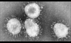 ration. But by late 1980’s her passion of electron microscopy led her back to ST. Thomas in advisory role where she with her colleagues published some high-quality negative staining electron micrographs of the Human Immunodeficiency Virus (HIV).
Before she passed away in 2007 she continued to image viruses, she had also co-published a document for World Health Organisation in 1979 titled “Manual For Rapid Laboratory Viral Diagnosis.